


Fibre study gives 3D view
 Print
Print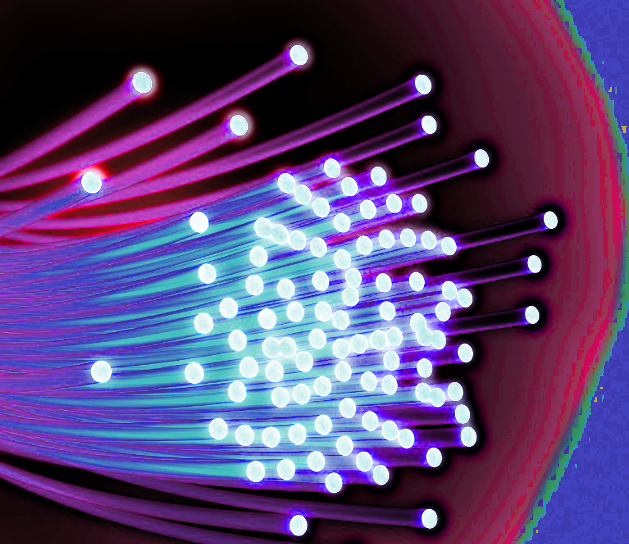 A new technique could see normal optical fibre technology used to produce microscopic 3D images.
Unlike normal biopsies where tissue is harvested and sent off to a lab for analysis, optical biopsies enable clinicians to examine living tissue within the body in real-time.
This minimally-invasive approach uses ultra-thin microendoscopes to peer inside the body for diagnosis or during surgery, but normally produces only two-dimensional images.
But experts sat RMIT has developed a technique to access the 3D potential of existing microendoscope technology.
The new technique uses a light field imaging approach to produce microscopic images in stereo vision, similar to 3D glasses for watching films.
“Stereo vision is the natural format for human vision, where we look at an object from two different viewpoints and process these in our brains to perceive depth,” said research leader Dr Antony Orth.
“We’ve shown it’s possible to do something similar with the thousands of tiny optical fibres in a microendoscope.
“It turns out these optical fibres naturally capture images from multiple perspectives, giving us depth perception at the microscale.
“Our approach can process all those microscopic images and combine the viewpoints to deliver a depth-rendered visualisation of the tissue being examined – an image in three dimensions.
“The key observation we made is that the angular distribution of light is subtly hidden in the details of how these optical fibre bundles transmit light,” Dr Orth said.
“The fibres essentially ‘remember’ how light was initially sent in – the pattern of light at the other side depends on the angle at which light entered the fibre.”
With this in mind, the researchers developed a mathematical framework to relate the output patterns to the light ray angle.
“By measuring the angle of the rays coming into the system, we can figure out the 3D structure of a microscopic fluorescent sample using just the information in a single image,” said researcher Professor Brant Gibson.
“So that optical fibre bundle acts like a miniaturised version of a light field camera.
“The exciting thing is that our approach is fully compatible with the optical fibre bundles that are already in clinical use, so it’s possible that 3D optical biopsies could be a reality sooner rather than later.”