


New tool for deep view of mind's molecules
 Print
Print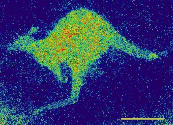 A new tool for imaging life at the nanoscale - developed in Australia - could provide new insights into the molecules involved in neuro-degenerative diseases.
In a new paper published today in Nature Communications, University of Melbourne researchers detected a custom-made reveal a “quantum kangaroo” in a demonstration of their new way to detect and image electronic spins non-invasively, and with a level of sensitivity and resolution orders of magnitude higher than previously achieved.
The breakthrough could one day help physicians and researchers investigate the role transition metal ions play in biology and disease.
Electron spin resonance (ESR) techniques have been used to understand biochemical processes in biological systems for quite some time, but there has not been same rapid growth in ESR technology that its sister technology, nuclear magnetic resonance (NMR), has.
NMR is the technology underpinning now-common magnetic resonance imaging (MRI).
Both ESR and NMR apply a magnetic field to image molecules, but unlike NMR, ESR can reveal biochemistry related to metal ions and free radicals.
In biological systems, the detectable concentration of electron spins is many orders of magnitude lower than nuclear spins. Hence, the roadblock for the development of ESR-based imaging techniques has been the sensitivity required – typically billions of electronic spins have been needed to generate a sufficient signal for successful imaging.
This is where quantum technology comes in.
A team led by Professor Lloyd Hollenberg has used a specially engineered array of quantum probes in diamond to demonstrate non-invasive ESR imaging with sub-cellular resolution. Remarkably, the system has been able to image and interrogate very small regions containing only a few thousand electron spins.
“The sensing and imaging technology we are developing enables us to view life in completely new ways, with greater sensitivity and resolution derived from the fundamental interactions of sample and probe at the quantum mechanical level,” said Dr Hollenberg.
“This dramatic improvement in ESR imaging technology is an exciting development and a clear demonstration of how quantum technology can be used to enhance signal sensitivity and provide solutions to long standing problems, for example probing human biochemistry at even finer scales.”
Scaling ESR technology down to sub-micron resolution has been challenging because such a reduction in spatial resolution requires substantially better sensitivity. However, this is precisely what quantum probes offer – high sensitivity with high spatial resolution.
By generating an array of quantum probes in diamond, using the material’s unique nitrogen-vacancy colour centre, the research team was able to detect and image electronic spin species at the diffraction limit of light, 300 nanometres.
Crucially, the sensing technology is able to provide spectroscopic information on the particular source of electronic spins being imaged.
“Transition metal ions are implicated in several neuro-degenerative diseases, however, little is known about their concentration and oxidation state within living cells,” said lead author Dr David Simpson.
“We aim to adapt this new form of sensing to begin probing such effects in a range of biological systems.”
One of the unique advantages of quantum-based sensing is that it does not interfere with the sample being imaged.
Other approaches rely on fluorescent molecules binding to particular targets of interest. While these approaches are species-specific, they modify the functionality and availability of the target species being imaged.
PhD student and co-author on the paper Robert Ryan explained the difference.
“Our technique relies on passive, non-invasive detection of electronic spins by observing their interaction with the quantum probe array,” Mr Ryan said.
“By carefully tuning an external magnet into resonance with the quantum probes, we are able to listen to the magnetic noise created by the sample’s electronic spins. Different electronic spin species have different resonance conditions; therefore we are able to detect and image various electronic spin targets.”